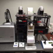
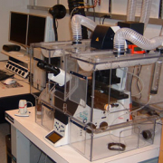
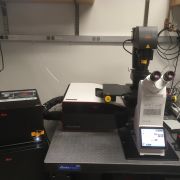
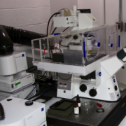
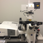
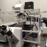
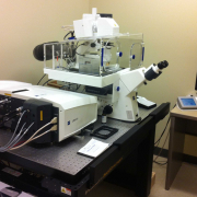
- High speed multi-modal imaging with optical sectioning with 25um and 40um pinhole sizes
- Four laser lines: 405nm, 488nm, 561nm, 637nm
- Environmental control for samples which need heating and CO2
- Microscope autofocus control for time-lapse imaging
- Additional technologies: TIRF, SRRF, point localization super-resolution, MicroPoint laser ablation, and Mosaic 3 photostimulation
- High-speed multidimensional imaging with optical sectioning
- Spinning disk modality produces high SNR images with minimal photobleaching
- High sensitivity EMCCD camera
- Laser lines for all classes of fluorscent proteins
- Incubation for mammalian cells
- IR reflection based autofocus
- Silicon oil 60x objective for RI match
Retired. Contact LMCF.
- High sensitivity point scanning confocal
- High-sensitivity GaAsP HyD detectors with photon counting
- Spectrally tunable emission band ranges
- Resonant scanner for low photo-toxicity
- Upright configuration
- 3D z-stacks, timelapse, stitching, multi-position timelapse
- Stimulated Emission Depletion super-resolution imaging
- High sensitivity point scanning confocal with pulsed white light laser
- High-sensitivity GaAsP HyD detectors with gating and photon counting
- Spectrally tunable emission bands
- Resonant scanner for low photo-toxicity
- Inverted configuration
- 3D z-stacks, timelapse, stitching, multi-position timelapse
- Leica point scanning confocal microscope
- Inverted Leica DMI8 microscope stand with Adaptive Focus Control and x,y scanning stage
- Pulsed White Light Laser (440nm-790nm) for imaging wide-range of fluorophores from blue to near infrared
- 5 high-sensitivity and low-noise spectral HyD detectors, all with photon counting and time-gating capability
- Imaging based on spectrum or by fluorescence lifetime: TauSense and Fluorescent Lifetime Imaging Microscopy (FLIM)
- Notch filters to further reduce background
- Conventional and resonant scanners
- LAS X software with 'LAS Dye Finder', 'LAS X 3D' image viewer and 'LAS X Navigator' for convenient tile-scanning and stitching
- Oko-Lab BoldLine 3 incubator allows multi-hour live cell imaging.
- High-resolution dual optical tweezers
- Three-color confocal fluorescence (488, 561, 638nm laser lines)
- Automated 5-channel microfluidic controls with dedicated laminar flow cell
- Imaging diverse fluorophores in fixed or live samples
- Temperature, humidity, CO2 control
- 3D z-stacks, timelapse, stitching, multi-position timelapse
- High sensitivity point scanning confocal imaging
- Spectral array GaAsP detectors
- Inverted microscope
- Incubation for live cell timelapse - temp and CO2 control
- Definite focus for timelapse
- High sensitivity point scanning confocal imaging
- Spectral array GaAsP detector
- Upright motorized stage microscope
- In vivo or in vitro samples
- FCS
- High sensitivity point scanning confocal imaging
- Spectral array GaAsP detectors
- AiryScan module with up to 2x resolution gain and high SNR (pdf)
- Fixed and live samples
- High sensitivity point scanning confocal imaging
- Spectral array GaAsP detectors
- AiryScan module with up to 2x resolution gain and high SNR (pdf)
- Fixed and live samples
- FCS and FCCS
