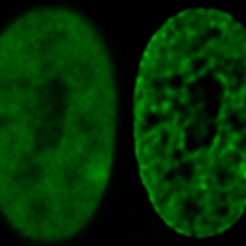
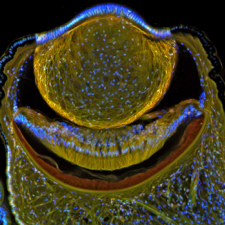
Menu

Secondary Menu

  • CoreResearch@Duke Login
  • News
logo
Light Microscopy Core Facility
  • About LMCF
    • Personnel
    • Location
    • Governance
    • Publications
    • Information for Publications
    • Funding Acknowledgements
    • Gallery of Images
  • Microscopes & Equipment
    • Microscopes
    • Image Analysis Workstations & Software
  • Services
    • Links to Image Analysis Software
    • Equipment Manuals
    • Guides & Tutorials
    • Data Saving/Data Transfer
  • Booking
    • New Users
    • Request Training
    • Fees
    • Facility Rules
    • CoreResearch@Duke Info
  • Feedback
  • Contact Us

Gallery of Images

 
arabidopsis embryo
Arabidopsis Embryo
 
muscle tendon
Muscle Tendon Morphogenesis
 
sea urchin embryo pharynx
Sea Urchin Embryo
Pharynx
 
confocal micrograph
Developing Drosophila
Egg Chamber
 
arabidopsis root
Arabidopsis Root
 
drosophila pupal morphogenesis
Drosophila Pupal
Morphogenesis
 
Podosomes
Podosomes
 
nanoprobe chip
Nanoprobe Chip
 
drosophila larval brain
Drosophila larval brain
 
lipid bilayer and monolayer
Lipid Bilayer and
Monolayer
 
butterfly nuclei
Butterfly Nuclei
 
sea urchin embryo
Sea Urchin Embryo
 
liver section
Liver Section
 
neonatal mouse cerebellum
Neonatal Mouse
Cerebellus
 
digestive system of wandering third instar larva
Digestive System
of Instar Larva
 
ovarian cancer cells
Ovarian Cancer Cells
 
dorsal closure
Dorsal Closure
in Drosophilia
 
beadring
Beadring
 
fibroblast
Podosomes and
Adhesion Complexes
 
pancreatic islet
Pancreatic Islet
 
mouse gut
3D Rendering of
Mouse Gut
 
tubulogenesis
Tubulogenesis
 
isosurface rendering
Isosurface Rendering
of Gene Locations
 
Cardiac progenitor cell cultures on PCL nanofibers
Cardia Progenitor Cell
Cultures on Nanofibers
 
mammalian cell nuclear
Mammalian Cell
Nuclear
 
skeletal C2C12 myoblasts
Skeletal Myoblasts
 
sea scallop
Eye of the Sea Scallop

Sidebar Navigation

  • Personnel
  • Location
  • Governance
  • Publications
  • Information for Publications
  • Funding Acknowledgements
  • Gallery of Images

Light Microscopy Core Facility (LMCF)

4215 French Family Science Center
124 Science Drive
Durham, NC 27708

lmcf@duke.edu
(919) 660.1234

Facility Operations

New Users
Reserve Equipment
Reservation Limits
Rules of Operation

Contacts & Logistics

Facility Access
Facility Locations
Feedback
Personnel
Report a Problem
CoreResearch@Duke Site

Services

Guides & Tutorials
Image Analysis
Manuals

Microscopes

Confocal Microscopes
Live Cell Imaging
Laser Capture Microdissection
Lightsheet Microscopes
Multiphoton
Stereoscopes & Dissecting
Super-resolution
TIRF
Widefield Fluorescence
NetID Login
  • About LMCF
    • Personnel
    • Location
      • French Family Science Cluster
      • Levine Science Research Center Cluster
      • Medical Sciences Research Building III Cluster
      • Nanaline Duke Cluster
    • Governance
    • Publications
    • Information for Publications
    • Funding Acknowledgements
    • Gallery of Images
  • Microscopes & Equipment
    • Microscopes
      • Confocal Microscopes
      • Live Cell Imaging
      • Laser Capture Microdissection
      • Lightsheet Microscopes
      • Multiphoton Microscopes
      • Stereoscopes & Dissecting
      • Super-resolution
      • TIRF
      • Widefield Fluorescence
    • Image Analysis Workstations & Software
  • Services
    • Links to Image Analysis Software
    • Equipment Manuals
      • Andor XD Spinning Disk Confocal Microscope Manual
      • Andor Dragonfly Spinning Disk Confocal Microscope (User's Manual)
      • DeltaVision Elite Deconvolution Microscope
      • Leica AM Total Internal Reflection Fluorescence Manual
      • Leica SP5 Inverted Confocal Manual
      • Live Cell Station (Zeiss Axio Observer Z1) Manual - With MetaMorph
      • Live Cell Station (Zeiss Axio Observer Z1) Manual - Single or Tile Imaging With ZEN
      • Olympus FV1000 Multiphoton Manual
      • Olympus VivaView FL Incubator Microscope Manual
      • ZEN 2.3 Pro: Single Field or Tiling/Stitching Multiple Fields
      • Zeiss 710 Inverted confocal manual
      • Zeiss 780 inverted confocal manual
      • Zeiss 780 upright confocal manual
      • Zeiss Axio Imager Z2 Upright Microscope Manual
      • Zeiss Axio Observer Manual
      • Zeiss LSM 510 Inverted Confocal Manual
      • Zeiss Lumar.V12 Stereoscope Manual
      • Zeiss MosaiX - Color brightfield tiling and image stitching on the LCM
      • Zeiss PALM MicroBeam Laser Capture Microdissection Manual
    • Guides & Tutorials
      • Introduction to Microscopy
      • Choose Best Microscope for your Sample
      • Introduction to Sample Preparation
        • Coverslip Thickness
        • Glass Coverslip Cleaning (Acid Wash)
        • Multiwell Chamber Slides
        • Paraformaldehyde, Formadehyde and Formalin
      • Safe Imaging of Live Samples
      • Laser Lines and Filters
      • Count Nuclear Foci - ImageJ
      • Overlay Images in ImageJ
      • Photoshop
      • Control Motorized Stage in MetaMorph
      • Kohler Illumination Procedure
      • Clean an Objective
      • File Formats for Microscopy
      • References
    • Data Saving/Data Transfer
  • Booking
    • New Users
    • Request Training
    • Fees
    • Facility Rules
      • Facility Access
      • Reservation Limits
    • CoreResearch@Duke Info
      • How to Make a Reservation
      • How to Change a Reservation
      • How to Record Actual Use
      • Tips for Using CoreResearch@Duke
  • Feedback
  • Contact Us
  • CoreResearch@Duke Login
  • News