- High speed multi-modal imaging with optical sectioning with 25um and 40um pinhole sizes
- Four laser lines: 405nm, 488nm, 561nm, 637nm
- Environmental control for samples which need heating and CO2
- Microscope autofocus control for time-lapse imaging
- Additional technologies: TIRF, SRRF, point localization super-resolution, MicroPoint laser ablation, and Mosaic 3 photostimulation

- Stimulated Emission Depletion super-resolution imaging
- High sensitivity point scanning confocal with pulsed white light laser
- High-sensitivity GaAsP HyD detectors with gating and photon counting
- Spectrally tunable emission bands
- Resonant scanner for low photo-toxicity
- Inverted configuration
- 3D z-stacks, timelapse, stitching, multi-position timelapse

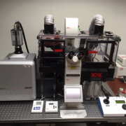
- Optical trapping of beads or protein or membrane complexes
- Confocal imaging between two optical traps with 488nm and 561nm lasers
- DNA–binding protein experiments: Study molecular mechanisms involved in DNA organization, repair, replication, transcription, and RNA translation.
- Others: Protein folding; Cytoskeleton structure and transport; Mechanobiology.
- Phase separation.
- Immuno-oncology.
- High sensitivity point scanning confocal imaging
- Spectral array GaAsP detectors
- AiryScan module with up to 2x resolution gain and high SNR (pdf)
- Fixed and live samples
- High sensitivity point scanning confocal imaging
- Spectral array GaAsP detectors
- AiryScan module with up to 2x resolution gain and high SNR (pdf)
- Fixed and live samples
- FCS and FCCS
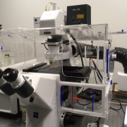
The ZEISS Elyra 7 is a versatile super-resolution microscope system which combines high-speed live-cell imaging with subcellular detail, offering resolution of 100-120nm with SIM and even down to 60 nm with Lattice SIM² or 20-30 nm with SMLM - single molecule localization microscopy (like PALM/dSTORM) for molecular precision. Its key capabilities include fast 3D imaging with dual sCMOS cameras, gentle illumination for live samples, wide-field capabilities, DIC, and TIRF or HILO.
Highly inclined and laminated optical sheet (HILO) and total internal reflection illumination (TIRF).
Apotome 2: Grid-based optical sectioning to create highly contrasted images with high lateral and axial resolution.