Shared resource offers efficient and low-cost access to advanced imaging technology
Department: Light Microscopy Core Facility
Years at Duke: 7
Number of employees: 3
Who they are: The Light Microscopy Core Facility is a shared scientific resource for Duke faculty, staff and students. Researchers can use any of nearly two dozen different high-tech imaging machines, from simple fluorescence microscopes to $500,000 confocal microscopes that create crisp optical slices of the sample. The Office of the Provost, School of Medicine, Duke Cancer Institute and grants fund the facility. Researchers from any department at Duke University and Duke University Health System can sign up to use machines for hourly fees that range between around $5 and $40 per hour, depending on the equipment. "We provide the machines and guidance on how to use them so researchers can carry out the imaging they need," said Sam Johnson, director of the facility.
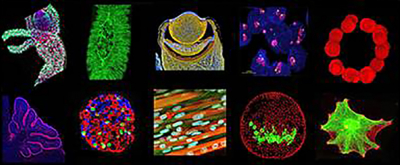
What they're known for: Obtaining images that help scientists understand the research topics they are working on. The images range enormously in topic - from a close-up of the eye of a sea scallop to the arrangement of fluorescent beads a hundredth the size of a grain of sand in response to a magnetic field.
Hidden department fact: In addition to offering individual help to researchers setting up experiments and imaging processes, the facility also offers group instruction. Approximately 250 people have taken a microscopy course at Duke to learn more about how the machines work, what they are capable of doing and how best to process and analyze the resulting images.
Significant achievement: The Light Microscopy Core Facility has significantly increased the available microscopy infrastructure at Duke. Thanks to institutional funding and grants from the National Institutes of Health and the North Carolina Biotechnology Center, Duke now has a wide range of equipment available to all researchers. "When we started building the microscopy facility seven years ago, the scopes were being used about 200 hours a month," Johnson said. "Now we've added many systems and users and the scopes are often used over 2,500 hours a month."
What they can do for you: "If you have a research sample that fluoresces or interacts with light in some way, we can probably help you image it or extract information from it," Johnson said. "We help people look at research samples with techniques that involve expensive equipment or specifc expertise or both."
Big goal: To provide access to the imaging techniques that researchers need, in adequate capacity in a way that is practical, efficient, convenient and affordable.
How they make a difference: "At Duke you no longer have to buy very expensive equipment or be an expert in microscopy in order to use the equipment," Johnson said. "We hope we lower the barriers for using imaging as part of research."